Paper – FTIR microscopy with RMieS
FTIR microscopy of biological cells and tissue: data analysis using resonant Mie scattering (RMieS) EMSC algorithm
Paul Bassan, Ashwin Sachdeva, Achim Kohler, Caryn Hughes, Alex Henderson, Jonathan Boyle, Jonathan H. Shanks, Michael Brown, Noel W. Clarke and Peter Gardner
Analyst 137 (2012) 1370-1377
Abstract
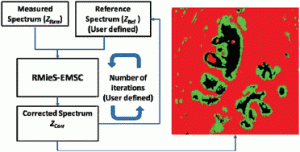
Transmission and transflection infrared microscopy of biological cells and tissue suffer from significant baseline distortions due to scattering effects, predominantly resonant Mie scattering (RMieS). This scattering can also distort peak shapes and apparent peak positions making interpretation difficult and often unreliable. A correction algorithm, the resonant Mie scattering extended multiplicative signal correction (RMieS-EMSC), has been developed that can be used to remove these distortions. The correction algorithm has two key user defined parameters that influence the accuracy of the correction. The first is the number of iterations used to obtain the best outcome. The second is the choice of the initial reference spectrum required for the fitting procedure. The choice of these parameters influences computational time. This is not a major concern when correcting individual spectra or small data sets of a few hundred spectra but becomes much more significant when correcting spectra from infrared images obtained using large focal plane array detectors which may contain tens of thousands of spectra. In this paper we show that, classification of images from tissue can be achieved easily with a few (<10) iterations but a reliable interpretation of the biochemical differences between classes could require more iterations. Regarding the choice of reference spectrum, it is apparent that the more similar it is to the pure absorption spectrum of the sample, the fewer iterations required to obtain an accurate corrected spectrum. Importantly however, we show that using three different non-ideal reference spectra, the same unique correction solution can be obtained.
