Imaging and pathology
Infrared spectral imaging of prostate cancer biopsies: Towards spectroscopic pathology.
Prostate cancer (CaP) is the second most common cause of cancer related death in males within the United Kingdom. At present, a prognosis for CaP is determined by its accurate assessment of disease grade and stage. To this end, the universally accepted method in histopathological grading is through the use of the Gleason grading system.

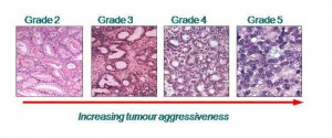
This method in grading sub-divides the complex continuum of glandular architectural changes that occur during prostate cancer progression into five visually distinct grades that can be viewed under an optical microscope. However, there are limitations in reproducibility in a specific grade due to intra and inter observer variability. These preclude its acceptance as a single prognostic tool. Methods to increase the accuracy of Gleason scoring by pathologists have been suggested and these rely on standardising the visual criterion of pattern recognition on which the system is currently based. In this project in collaboration with Noel Clark at the Paterson Institute for Cancer Research and Jonathan Shanks at the Christie Hospital in Manchester we have used infrared spectroscopy and spectroscopic imaging to study prostate cancer biopsies. The aim of this project is to develop a robust and reliable method of tissue analysis that will augment the current imunohistochemical methods currently employed by pathologist in order to aid faster and more accurate diagnosis. In a pilot study we have shown that it is possible to grade the tissue spectroscopically and this opens up the possibility of developing an automated system of diagnosis. We have also shown that spectra can be use to determine the stage of the disease.

Publications related to this project
Investigating FTIR Based Histopathology for the Diagnosis of Prostate Cancer
M.J. Baker, E. Gazi, M.D. Brown, J.H. Shanks, N.W. Clarke, P. Gardner
J. Biophotonics, 2 (2009) 104-113
FTIR Based Spectroscopic Analysis in the Identification of Clinically AggressiveProstate Cancer
M.J. Baker, E.Gazi, M.D. Brown, J.H. Shanks, P.Gardner, N.W. Clarke
British Journal of Cancer, 99 (2008) 1859-1866
A Correlation of FTIR Spectra Derived from Prostate Cancer Tissue with Gleason Grade, and Tumour Stage
E. Gazi, M. Baker, J. Dwyer, N. P. Lockyer, P.Gardner, J.H. Shanks, R. S. Reeve, C. Hart, N.W. Clarke M. Brown
European Urology 50 (2006) 750–761
Application of FTIR Microspectroscopy and ToF-SIMS Imaging in the Study of Prostate Cancer
E. Gazi, J. Dwyer, N. Lockyer, P. Gardner, J.C.Vickerman, J. Miyan, C. Hart, M. Brown and N. Clarke
Faraday Discussions 126 (2004) 41 – 59
Applications of Fourier Transform Infrared Microspectroscopy in Studies of Benign Prostate & Prostate Cancer. A Pilot Study
E. Gazi, J. Dwyer, P. Gardner, A.Ghanbari-Siakhali, A. P. Wade. J. Myan. N.P.Lockyer, J. C. Vickerman, N. W.Clarke, J. H. Shanks, C. Hart, M.Brown
J. Pathology 201 (2003) 99-108
Currently working on this project
Konrad Dorling
Funding
- EPSRC/RSC Analytical Studentship
- Williamson Trust